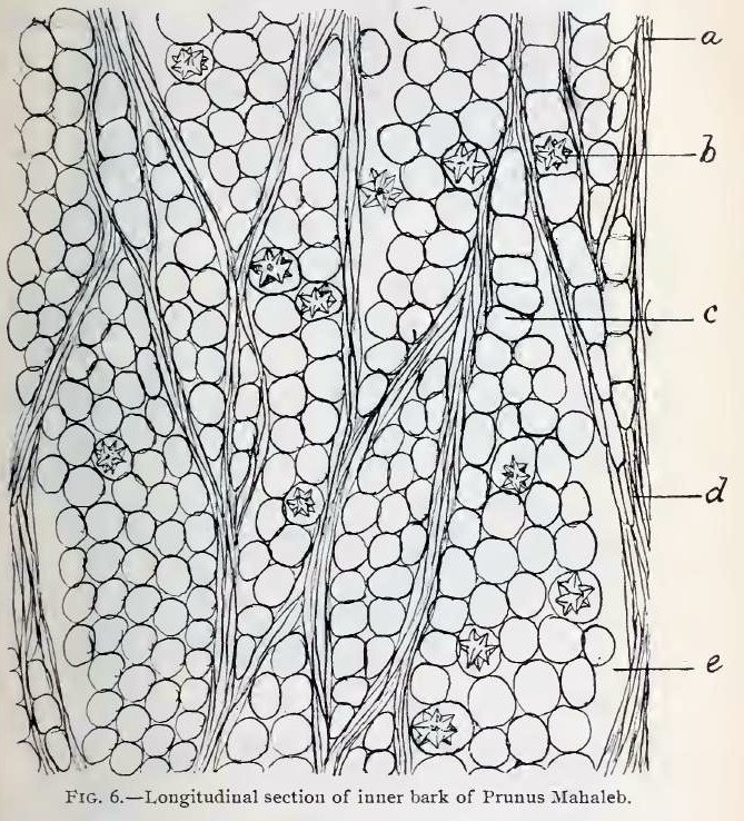
Fig. 6.—Tangential section through bast layer of Prunus Mahaleb, showing medullary rays and compressed sieve tissue. Magnification about 230 diameters.
a, compressed sieve tissue;
b, crystal cell;
c, ordinary parenchyma cell of medullary ray;
d, compressed sieve tissue;
e, fissure between medullary ray and sieve tissue.
This image is from Structure of our Cherry Barks in the September issue of the American Journal of Pharmacy, 1895.
